Description
An enzyme immunoassay for the qualitative and semiquantitative determination of IgA-class antibodies to Adenovirus in serum.
Adenoviruses are double-stranded DNA viruses lacking an envelope, of about 60-90 nm. The capsid contains 252 capsomeres and shows icosahedral symmetry. The capsomeres
consist of hexons, pentons, and fibers (so named after their configuration) which are responsible for the induction of group- and type-specific antibodies. In humans, 34 immunologically distinct types (serotypes) are recognized. Adenoviruses were first isolated from adenoid tissue and have a certain affinity for lymph glands, where they may remain latent for years. They also invade the respiratory tract, the gastrointestinal tract, and the conjunctiva. Adenovirus infections are widely distributed and common with most infections occurring in childhood. The contagious disease normally is acute and self-limited, but infections may be prolonged and asymptomatic, possibly remaining latent for a very long time. Winter and Spring are the seasons for the highest rates of general adenovirus infections, independent of geographic locations and climatic conditions. Epidemics may occur in over-crowded populations, for example ARD in military groups, PCF in swimming pools, and EKC in medical facilities.
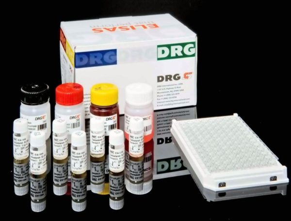
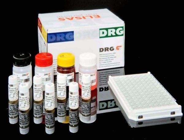
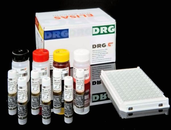
The DRG Adenovirus IgA ELISA Kit is a solid phase enzyme-linked immunosorbent assay (ELISA) Microtiter wells as a solid phase are coated with Adenovirus antigen. Patient
samples are diluted with Sample Diluent and additionally incubated with IgG-RF-Sorbent, containing hyper-immune anti-human IgG-class antibody to eliminate competitive
inhibition from specific IgG and to remove rheumatoid factors. This pretreatment avoids false negative or false positive results.Diluted patient specimens and ready-for-use controls are pipetted into these wells. During incubation Adenovirus-specific antibodies of positive specimens and controls are bound to the immobilized antigens. After a washing step to remove unbound sample and control material horseradish peroxidase conjugated anti-human IgA antibodies are dispensed into the wells. During a second incubation this anti_IgA conjugate binds specifically to IgA antibodies resulting in the formation of enzyme-linked immune complexes. After a second washing step to remove unbound conjugate the immune complexes formed (in case of positive results) are detected by incubation with TMB substrate and development of a blue color. The blue color turns into yellow by
stopping the enzymatic indicator reaction with sulfuric acid. The intensity of this color is directly proportional to the amount of Adenovirus-specific IgA antibody in the patient
specimen. Absorbance at 450 nm is read using an ELISA microtiter plate reader.


