Description
An enzyme immunoassay for the qualitative and semiquantitative determination of IgM-class antibodies to Candida albicans in serum.
Species of Candida are non-mycelia-producing non-ascospore-forming yeast-like fungi which appear as small (4-6 µm), oval, thin-walled gram-positive budding cells. The most
important representative, Candida albicans, is a facultative pathogen for man. C. albicans is ubiquitous and commonly found as transient flora on normal mucous membranes. Although not pathogenic in healthy humans the fungus may be opportunistic in those suffering from a variety of disorders, and in those treated intensively with broad-spectrum antibiotics or immunosuppressive measures. Candidiasis is caused about 90% of the time by C. albicans. It is an acute or subacute infection in which the fungus may produce lesions in the mouth (thrush, oral candidiasis), vagina (vulvovaginal candidiasis), skin and nails (intertriginous candidiasis), bronchi or lungs (bronchopulmonary candidiasis) and, occasionally, a septicemia, endocarditis or meningitis. In immunosuppressant patients with cellular immunodeficiency, e.g., AIDS patients, C. albicans may lead to severe necrosis
of infected tissues.
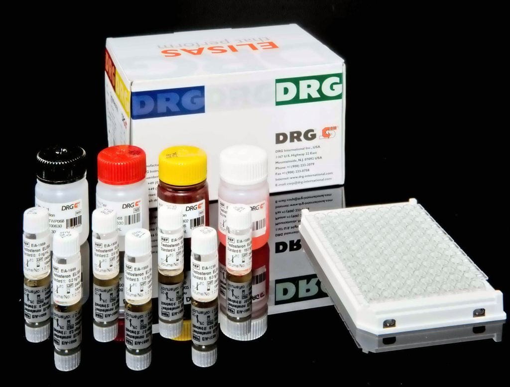
The DRG Candida albicans IgM ELISA Kit is a solid phase enzyme-linked immunosorbent assay (ELISA) Patient samples are diluted with Sample Diluent and additionally incubated with IgG-RF-Sorbent, containing hyper-immune anti-human IgG-class antibody to eliminate competitive inhibition from specific IgG and to remove rheumatoid factors. This pretreatment avoids false negative or false positive results. Microtiter wells as a solid phase are coated with Candida albicans antigen.Pretreated patient specimens and ready-for-use controls are pipetted into these wells. During incubation Candida albicans-specific antibodies of positive specimens and controls are bound to the immobilized antigens. After a washing step to remove unbound sample and control material horseradish peroxidase conjugated anti-human IgM antibodies are dispensed into the wells. During a second incubation this anti_IgM conjugate binds specifically to IgM antibodies resulting in the formation of enzyme-linked immune complexes. After a second washing step to remove unbound conjugate the immune complexes formed (in case of positive results) are detected by incubation with TMB substrate and development of a blue color. The blue color turns into yellow by stopping the enzymatic indicator reaction with sulfuric acid. The intensity of this color is directly proportional to the amount of Candida albicans-specific IgM antibody in the patient specimen. Absorbance at 450 nm is read using an ELISA microtiter plate reader.


