Description
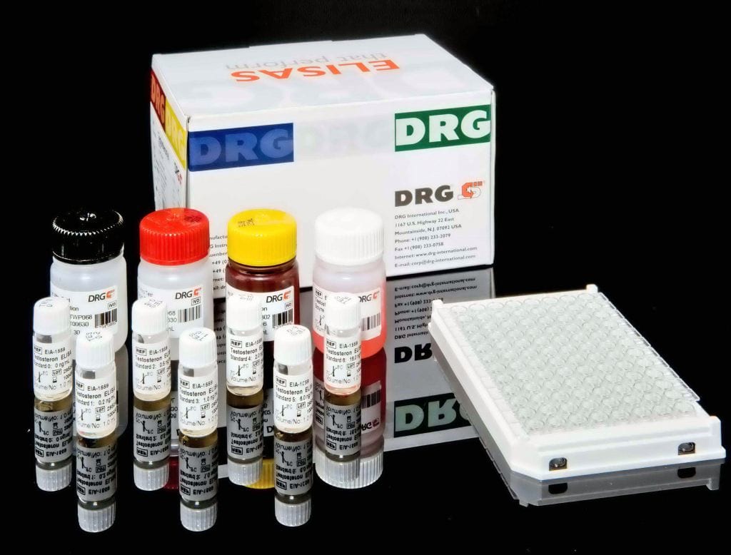
An enzyme immunoassay for the quantitative determination of carcinoembryonic antigen (CEA) in serum.
Carcinoembroyonic antigen (CEA) is a cell-surface 200-kd glycoprotein. In 1969, it was reported that plasma CEA was elevated in 35 of 36 patients with adenocarcinoma of the colon and that CEA titers decreased after successful surgery. Normal levels were observed in all patients with other forms of cancer or benign diseases. Subsequent studies have not confirmed these initial findings, and it is now understood that elevated levels of CEA are found in many cancers. Increased levels of CEA are observed in more than 30% of patients with cancer of the lung, liver, pancreas, breast, colon, head or neck, bladder, cervix, and prostate. Elevated plasma levels are related to the stage and extent of the disease, the degree of differentiation of the tumor, and the site of metastasis. CEA is also found in normal tissue.
The CEA ELISA test is based on the principle of a solid phase enzyme-linked immunosorbent assay. The assay system utilizes a monoclonal antibody directed against a distinct antigenic determinant on the intact CEA molecule is used for solid phase immobilization (on the microtiter wells). A goat anti-CEA conjugated to horseradish peroxidase (HRP) is in the antibody-enzyme conjugate solution. The test sample is allowed to react simultaneously with the two antibodies, resulting in the CEA molecules being sandwiched between the solid phase and enzyme-linked antibodies. After a 1 hour incubation at room temperature, the wells are washed with water to remove unbound labeled antibodies. A solution of TMB Reagent is added and incubated for 20minutes, resulting in the development of a blue color. The color development is stopped with the addition of Stop Solution changing the color to yellow. The concentration of CEA is directly proportional to the color intensity of the test sample. Absorbance is measured spectrophotometrically at 450 nm.


