Description
An enzyme immunoassay for the quantitative and qualitative determination of IgA-class antibodies to Helicobacter pylori in serum and plasma.
Helicobacter pylori is a spiral Gram-negative bacterium (2-6.5 µm in size, flagellated) which colonizes the human gastric mucosa. The organism is found in the mucus layer and adheres to the surface mucus epithelium of the stomach but generally does not penetrate the gastric mucosa directly. However, there is a secondary inflammatory response in the mucosa leading to chronic active gastritis. Helicobacter pylori is the primary causative agent in most cases of peptic ulcer disease. In 1994 the WHO classified Helicobacter pylori as a category 1 carcinoma. Infection rate in Europe is about 30%-40%, worldwide about 50%. There is an inverse relationship between the presence of Helicobacter pylori infection and socioeconomic status. In developing countries, people acquire the infection at an early age such that by young adulthood as many as 90% of the population might have Helicobacter pylori gastritis. In developed western countries the prevalence of Helicobacter pylori gastritis is much lower. Under these conditions, the rate of acquisition is much slower (roughly 1% per annum) and the older one is, the more likely one is to be infected with the organism.
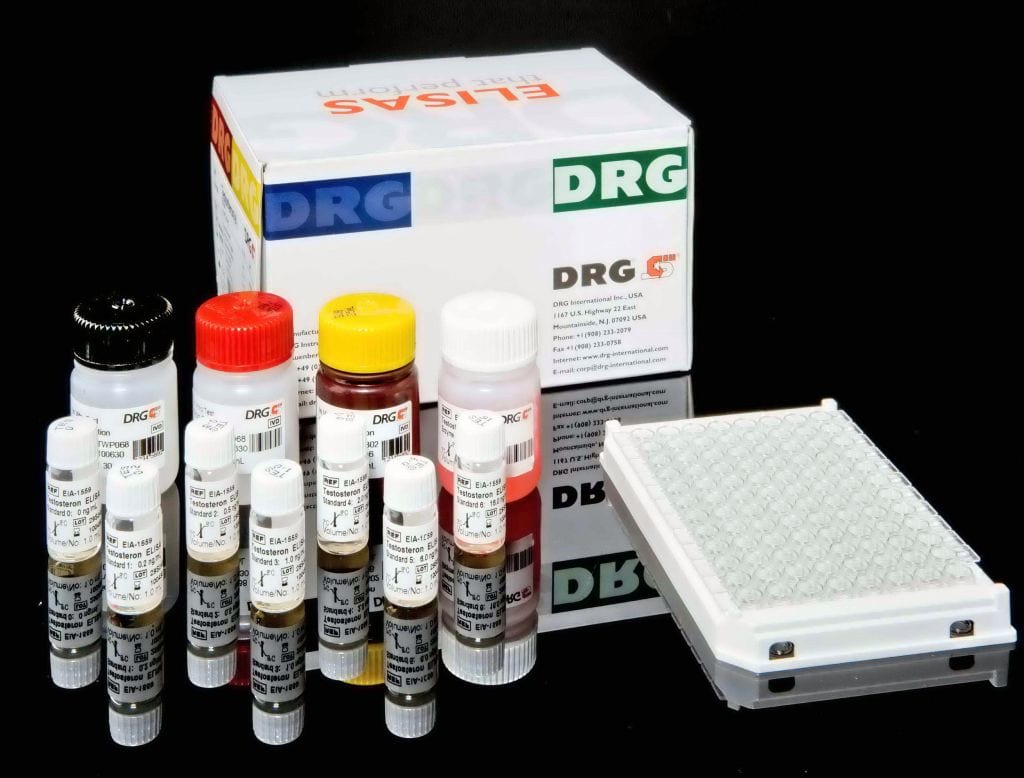
The DRG Helicobacter pylori IgA ELISA Kit is a solid phase enzyme-linked immunosorbent assay (ELISA)Patient samples are diluted with Sample Diluent and additionally incubated with IgG-RF-Sorbent, containing hyper-immune anti-human IgG-class antibody to eliminate competitive inhibition from specific IgG and to remove rheumatoid factors. This pretreatment avoids false negative or false positive results. Microtiter wells as a solid phase are coated with inactivated recombinant Helicobacter pylori (CagA) antigen. Diluted patient specimens and ready-for-use controls are pipetted into these wells. During incubation Helicobacter pylori-specific antibodies of positive specimens and controls are
bound to the immobilized antigens. After a washing step to remove unbound sample and control material horseradish peroxidase conjugated anti-human IgA antibodies are dispensed into the wells. During a second incubation this anti_IgA conjugate binds specifically to IgA antibodies resulting in the formation of enzyme-linked immune complexes. After a second washing step to remove unbound conjugate the immune complexes formed (in case of positive results) are detected by incubation with TMB substrate and
development of a blue color. The blue color turns into yellow by stopping the enzymatic indicator reaction with sulfuric acid. The intensity of this color is directly proportional to the amount of Helicobacter pylori-specific IgA antibody in the patient specimen. Absorbance at 450 nm is read using an ELISA microtiter plate reader.


