Description
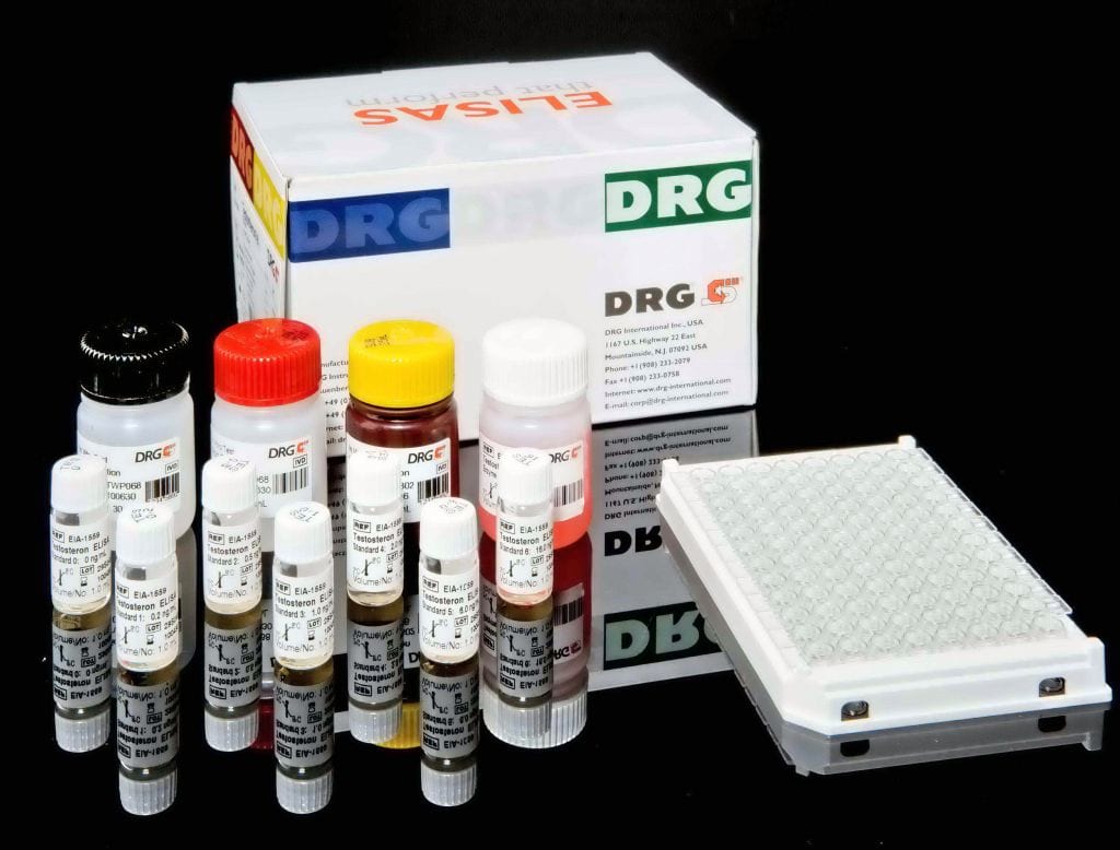
An enzyme immunoassay for the quantitative determination of free prostate specific antigen (f-PSA) in serum or plasma.
Prostate cancer is the most frequent type of cancer found in man and is the second cause of death due to cancer in males. Until recently, digital rectal examination (DRE) was frequently used as only diagnostic modality for the detection of early stages of prostate cancer. In the recent years the determination of serum PSA levels has become the most accepted method to improve the diagnostic specificity of DRE. Although PSA is a tissue specific protein and is not solely tumor specific, it has become the most important marker
for prostate carcinoma, showing a better specificity than other biochemical markers used in this context (PAP, total alkaline phosphatase, carcinoembryonic antigen, etc.) In 1979, Wang et al. isolated a specific antigen for normal prostate tissue and called this protein PSA. PSA is a 33 kDa serine proteinase. Immunohistological studies have shown that PSA is localized in the cytoplasm of prostate acinar cells, ductal epithelium and in the secretion on the ductal lumina, present in normal, benign hyperplastic and malignant prostate tissues
as well metastatic prostate cancer and in seminal plasma. If the structural integrity of the prostate is disturbed and/or the gland size is increased, the amount of PSA in the blood plasma may become elevated. In the blood plasma, most of the PSA forms complexes with various proteinase inhibitors. Only a small fraction of PSA circulates as free inactive PSA. Basically three major forms of PSA can be distinguished, only two of which are immunoreactive. The predominant form of PSA is a complex with _1-antichymotrypsin (ACT-PSA).
Inactive free PSA (f-PSA) represents around 10-40% of the immunologically detectable PSA. The total amount of immunoreactive PSA is known as total PSA (t-PSA). PSA complexed with _- 2-macroglobulin cannot be detected by immunological assays and is therefore frequently called occult PSA (o-PSA). Current methods of screening men for prostate cancer utilize the detection of t-PSA. Levels of 4.0 ng/ml or higher are strong indicators of the possibility of prostatic cancer and are an indication for follow-up
examinations of the patient. However, elevated serum PSA levels are frequently also attributed to benign prostatic hyperplasia, leading to a high percentage of false positive screening results. A potential solution to this problem involves the determination of free PSA levels. Studies have suggested that the percentage of free PSA is lower in patients with prostate cancer than those with benign prostatic hyperplasia. Thus, the measurement of free serum PSA in conjunction with total PSA, can improve specificity of prostate cancer
screening in selected men with elevated total serum PSA levels, which would subsequently reduce unnecessary prostate biopsies with minimal effects on cancer detection rates.
This f-PSA ELISA is a solid phase enzyme-linked immunosorbent assay (ELISA). The microtiter wells are coated with an anti-f-PSA monoclonal antibody, directed towards an epitope of an antigen molecule. An aliquot of patient serum is incubated in the coated well with enzyme conjugated second antibody (E-Ab), directed towards a different region of the antigen molecule. After incubation the unbound E-Ab is washed off: The amount of bound E-Ab is proportional to the concentration of antigen in the sample. After adding the
substrate solution, the intensity of colour developed is proportional to the antigen concentration in the sample. The measured ODs of the standards are used to construct a calibration curve against which the unknown samples are calculated.


