Description
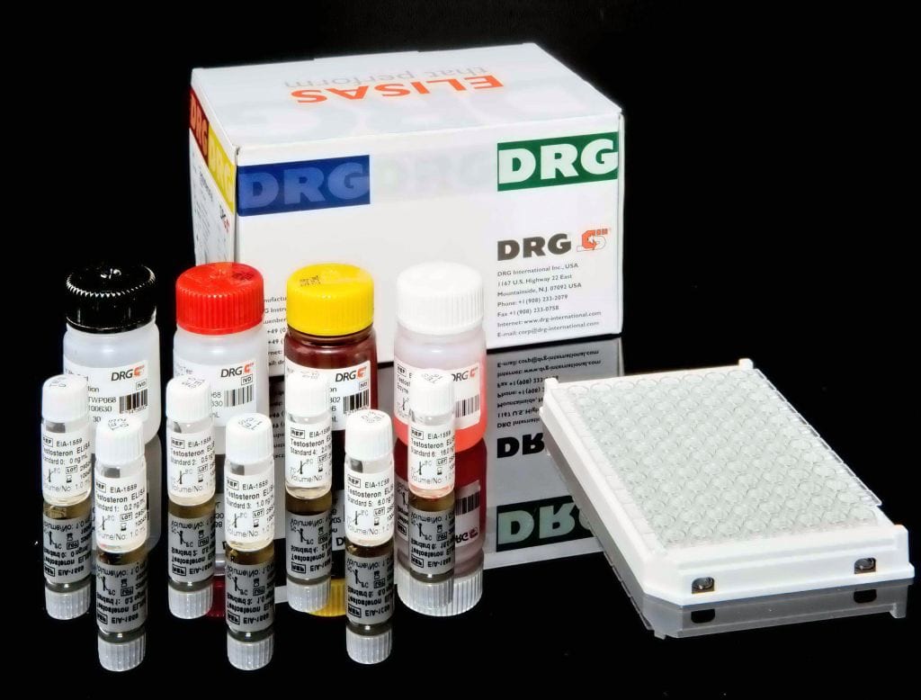
Rheumatoid Factor Screen ELISA is a test system for the quantitative measurement of IgG, IgM and IgA class rheumatoid facto in human serum or plasma.
This product is intended for professional in vitro diagnostic use only.
Fc fragments of highly purified human Immunoglobulin G are coated on to microwells. The determination is based on an indirect enzyme linked immune reaction with the following steps:
Specific antibodies in the patient sample bind to the antigen coated on the surface of the reaction wells. After incubation, a washing step removes unbound and unspecifically bound serum or plasma components. Subsequently added enzyme conjugate binds to the immobilized antibody-antigen-complexes. After incubation, a second washing step removes unbound enzyme conjugate. After addition of substrate solution the bound enzyme conjugate hydrolyses the substrate forming a blue coloured product. Addition of an acid stops the reaction generating a yellow end-product. The intensity of the yellow colour correlates with the concentration of the antibody-antigen-complex and can be measured photometrically at 450 nm.
The presence of IgM rheumatoid factor (RF) in the serum is the sole serological indicator included in the ACR list of criteria for the diagnosis of RA. RFs are a subset of anti globulins directed against the Fc region of IgG. In this definition we do not include antibodies to the IgG Fab region and pepsin agglutinators, directed against neo-antigens on IgG
exposed by pepsin cleavage. It is claimed that the majority of anti-globulin activity in normal serum is Fab-specific, whereas anti-globulin from RA patients is mostly Fc-specific. RFs are present in the serum of 75-80% of patients with RA at some time during the disease course. However, RFs are also found in the serum of patients with infectious and autoimmune diseases, hyperglobulinemia, B-cell lymphoproliferative disorders and in the aged population. This suggests that RF may be a finding associated with B-cell hyperactivity. Rheumatoid factors which have been found among the IgM, IgG and IgA classes of immunoglobulins, reacting only with xenogeneic Fc are not autoantibodies and are unlikely to be of pathological significance. However, RFs can bind IgG from many species, including autologous IgG, when immobilised on surfaces. Autologous binding is of higher affinity than xenogeneic binding. The here presented test systems for the determination of rheumatoid factors uses only human Fc fragments as coated antigen. It is generally considered that high titers of RF are associated with more severe disease and the presence of extra-articular features and rheumatoid nodules. This conclusion may depend on the disease duration. Serum IgM RF may precede the onset of RA by several years. A high titer of RF in non-RA individuals is associated with increased risk of developing RA. In the first 2 years of RA (early RA), serum levels of IgM, IgG and IgA RF do not correlate with disease activity. Serum IgG and IgA RF in these years are prognostic of erosive joint disease. In established RA, high titer serum IgM RF correlates with the presence of articular disease and nodules but not with systemic disease activity. The presence of either IgG or IgA RF in patients with long-standing RA may be a good prognostic indicator of systemic manifestations. IgG and IgM RF are associated with extra-articular RA including
rheumatoid vasculitis and nodules. The presence of IgM RF containing immune complexes with bound complement (C1q) C is also associated with extra-articular RA.


