Description
An enzyme immunoassay for the qualitative and semiquantitative determination of IgA-class antibodies to Respiratory Syncytial Virus (RSV) in serum.
Respiratory syncytial virus (RSV) is a negative-sense, enveloped RNA virus. The virion is variable in shape and size (average diameter of between 120 and 300 nm), is unstable in the environment (surviving only a few hours on environmental surfaces), and is readily inactivated. It is the causative pathogen for the most common infection of the respiratory tract.
Respiratory syncytial virus (RSV) infections usually occur during annual community outbreaks during the late fall, winter, or early spring months. The timing and severity of outbreaks in a community vary from year to year. Most infants become infected with RSV in their first winter season; between 25% and 40% have signs or symptoms of bronchiolitis or pneumonia, and 0.5% to 2% require hospitalization. Most children recover from illness in 8 to 15 days; the majority of children hospitalized for RSV infection are under 6 months of age. Most children will have serological evidence of RSV infection by 2 years of age. RSV also causes repeated infections throughout life, usually associated
with moderate-to-severe cold-like symptoms; however, severe lower respiratory tract disease may occur at any age, especially among elderly or among those with compromised cardiac, pulmonary, or immune systems.
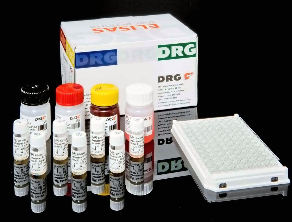
The DRG RSV IgA ELISA Kit is a solid phase enzyme-linked immunosorbent assay (ELISA) Microtiter wells as a solid phase are coated with inactivated RSV antigen. Diluted patient specimens and ready-for-use controls are pipetted into these wells. During incubation RSV-specific antibodies of positive specimens and controls are bound to the immobilized antigens. After a washing step to remove unbound sample and control material horseradish peroxidase conjugated anti-human IgA antibodies are dispensed into the wells. During a second incubation this anti_IgA conjugate binds specifically to IgA antibodies resulting in the formation of enzyme-linked immune complexes. After a second washing step to remove unbound conjugate the immune complexes formed (in case of positive results) are detected by incubation with TMB substrate and development of a blue color. The blue color turns into yellow by stopping the enzymatic indicator reaction with sulfuric acid. The intensity of this color is directly proportional to the amount of RSV-specific IgA antibody in the patient specimen. Absorbance at 450 nm is read using an ELISA microtiter plate reader.


