Description
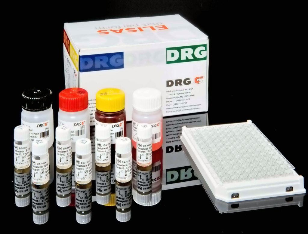
The human sVCAM-1 ELISA is an enzyme-linked immunosorbent assay for the quantitative detection of human sVCAM-1.The human sVCAM-1 ELISA is for in vitro diagnostic use. Not for use in therapeutic procedures.The vascular cell adhesion molecule-1 (VCAM-1) or CD106 is a member of the immunoglobulin gene superfamily. The initial molecular cloning of VCAM-1 reported six extracellular Ig-like domains (6D VCAM-1). This 6D VCAM-1 arises due to alternative splicing from a seven-domain VCAM-1 (7D VCAM-1). 7D VCAM-1 is the dominant form expressed by cultured human endothelial cells. Domains 1 through 3 are highly homologous to domains 4 through 6, suggesting that they arose by gene duplication. The cDNA of 7D VCAM-1 predicts a core protein of approximately 81 kDa with seven potential N-linked glycosylation sites. Upon complete glycosylation the mature protein has a molecular weight of approximately 102 kDa. This observation is in general agreement with immunoprecipitation studies that show a protein of approximately 110 kDa on cytokine-activated endothelium. Murine and rat VCAM-1 have been cloned. In contrast toICAM-1, VCAM-1 appears to have been highly conserved through evolution. Both rat and mouse VCAM-1 are highly homologous at the protein level to the human VCAM-1 (77% and 76%, respectively). VCAM-1 supports the adhesion of lymphocytes, monocytes, natural killer cells,eosinophils, and basophils through its interaction with leukocyte very late antigen-4 (VLA-4). VCAM-1/VLA-4 interaction mediates firm adherence of circulating non-neutrophilic leukocytes to endothelium. VCAM-1 also participates in leukocyte adhesion outside of the vasculature, mediating precursor lymphocyte adhesion to bone marrow stromal cells and B cell binding to lymph node follicular dendritic cells. VCAM-1 is not constitutively expressed on endothelium, but can be up-regulated in vitro in response to LPS, TNF-a, andIL-1, as well as to interferon-g and IL-4. VCAM-1 is also present on tissue macrophages, dendritic cells, bone marrow fibroblasts, myoblasts and myotubes. A soluble form of VCAM-1 (sVCAM-1) has been described. Soluble VCAM-1 levels have been found in the serum of healthy individuals and increased levels of sVCAM-1 can be detected in several diseases:
Cancer: ovarian, gastric-intestinal, renal, bladder cancer, non-Hodgkin’s lymphoma, breast cancer (cyst fluid);
Autoimmune diseases: multiple sclerosis (cerebrospinal fluid), systemic sclerosis, systemic lupus erythematosus, rheumatoid arthritis;
Infections: sepsis, meningitis, malaria;Inflammation: vasculitis, alcoholic cirrhosis, primary biliary cirrhosis, Wegener’s granulomatosis;
Others: impaired renal function,haemodialysis, hyperthyroidism, renal allograft.
Ananti-human sVCAM-1 coating antibody is adsorbed ontomicrowells.Human sVCAM-1present in the sample or standard binds to antibodies adsorbed to themicrowells. A conjugate mixture (biotin-conjugated anti-humansVCAM-1 antibody and Streptavidin-HRP) is added. Biotin-conjugatedanti-human sVCAM-1 antibody binds to human sVCAM-1captured by the first antibody.Streptavidin-HRPbinds to the biotin-conjugated anti-human sVCAM-1antibody.Followingincubation unbound Streptavidin-HRP is removed during a wash step, andsubstrate solution reactive with HRP is added to the wells.A coloured product is formed in proportion to theamount of human sVCAM-1 present in the sample orstandard. The reaction is terminated by addition of acid and absorbance ismeasured at 450 nm. A standard curve is prepared from 6 humansVCAM-1 standard dilutions and human sVCAM-1sample concentration determined.


