Description
The DRG:HYBRiD-XL free T3 is an enzyme immunoassay for the quantitative in vitro diagnostic measurement of free Triiodothyronine (T3) in serum and EDTA plasma.
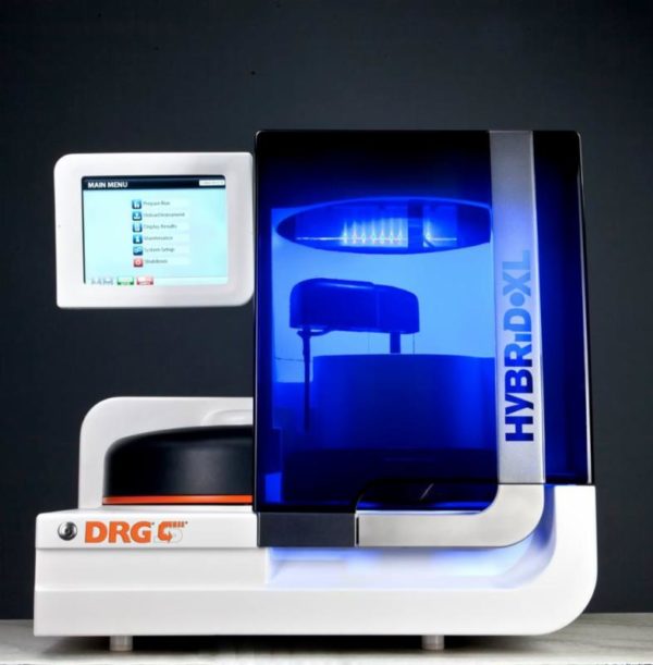
Only for use with the DRG:HYBRiD-XL Analyzer.
Triiodothyronine, also known as T3, is produced in the thyroid gland, the liver, the kidney and peripheral organs from its precursor thyroxine (T4). T3 affects almost every physiological process in the body, including growth and development, metabolism, body temperature, and heart rate. Production of T3 and T4 is induced by thyroid-stimulating hormone (TSH), which is released from the pituitary gland. More than 80% of T4 is produced by the thyroid gland, but only 15-20 % of T3 is produced there, while the rest is generated from T4 in liver, kidney and peripheral organs (1). More than 99.7% of T3 is bound to serum proteins, especially to Thyroxin binding globulin, but also to Transthyretin,
Albumin, and Lipoproteins (2,3). Since T3 has a 10-fold lower affinity to serum binding proteins than T4, free T3 concentrations are less influenced by variations of the concentration of thyroid hormone-binding proteins (4). In hyperthyroidism, free T3 concentrations are generally elevated. In hypothyroidism, free T3 concentrations tend to be lower, but the decrease is insufficient to give clear diagnostic information. Determination of free T3 is of clinical relevance for the detection of T3-hyperthyroidism and the early
stage or relapse of hyperthyroidism, and can be used for monitoring of Thyroxine treatment in hypothyroidism (6). free T3 determinations should be used as part of a thyroid test strategy, which may include free T4 and high sensitivity TSH tests.
The DRG:HYBRiD-XL free T3 Kit is a solid phase enzyme-linked immunosorbent assay (ELISA) based on the principle of competitive binding. The antibody coated wells (ACW) of the reagent cartridges are coated with a polyclonal [sheep] antibody directed towards a unique antigenic site of the T3 molecule. Endogenous free T3 of a –patient sample competes with a T3-horseradish peroxidase conjugate for binding to the coated antibody. After incubation the unbound conjugate is washed off. The amount of bound peroxidase conjugate is inversely proportional to the concentration of free T3 in the sample. Having added the substrate solution, the intensity of colour developed is inversely proportional to the concentration of free T3 in the patient sample.