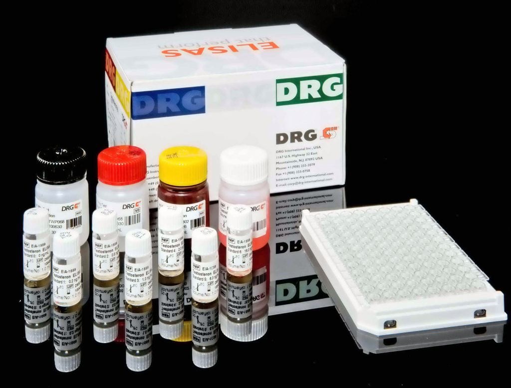
Description
Anti-Tissue-Transglutaminase IgG is an ELISA test system for the quantitative measurement of IgG class autoantibodies to tissue-transglutaminase (tTG) in human serum or plasma.
This product is intended for professional in vitro diagnostic use only.
Human recombinant tissue transglutaminase is bound to microwells. The determination is based on an indirect enzyme linked immune reaction with the following steps:
Specific antibodies in the patient sample bind to the antigen coated on the surface of the reaction wells. After incubation, a washing step removes unbound and unspecifically bound serum or plasma components. Subsequently added enzyme conjugate binds to the immobilized antibody-antigen-complexes. After incubation, a second washing step removes unbound enzyme conjugate. After addition of substrate solution the bound enzyme conjugate hydrolyses the substrate forming a blue coloured product. Addition of an acid stops the
reaction generating a yellow end-product. The intensity of the yellow color correlates with the concentration of the antibody-antigen-complex and can be measured photometrically at 450 nm.
Celiac disease (CD) comprises intolerance against dietary gluten present in wheat, rye and barley, and it belongs to the most common food-related diseases. Nowadays, CD is conceived as an autoimmune-mediated systemic disorder commonly presenting as enteropathy in genetically susceptible individuals. Four possible presentations of CD have been recognized: Typical, characterized mostly by gastrointestinal signs and symptoms. Atypical or extra intestinal, where gastrointestinal symptoms are minimal or absent and a number of other manifestations are present. Silent, where the small intestinal mucosa is damaged and CD autoimmunity can be detected by serology, but there are no symptoms. Latent, where individuals possess genetic compatibility with CD and may also show positive autoimmune serology, but have normal mucosa morphology and may or may
not be symptomatic.
The most obvious feature distinguishing CD from other small-intestinal enteropathies is the presence of autoantibodies against the key autoantigen tissue transglutaminase (abbreviated as TG2 or tTG) during a gluten containing diet. The gluten-derived gliadin peptides and the self antigen tTG, play a role in CD pathogenesis. Determination of serum levels of immunoglobulin A (IgA) against tTG is the first choice in suspected CD, displaying the highest levels of sensitivity and specificity. tTG is known to deamidate and crosslink gluten-derived gliadin peptides between a lysine and a glutamine residue. The interplay between gliadin peptides and tTG
is responsible for the generation of novel antigenic epitopes, the tTG-generated deamidated gliadin peptides (DGP). Such peptides represent much more CD-specific epitopes than native gliadin peptides, and anti-DGP antibodies are promising serological markers for CD.Endomysial antibodies (EMA) complement the repertoire of CD specific antibodies. EMA testing with indirect immunofluorescence methods may be a useful alternative if the result of the tTG test is equivocal. Tests for the detection of IgG or IgA antibodies against native gliadin peptides may be valuable to document adherence to the gluten-free diet administered by the
reating physician – the very effective but until now also the only possible treatment of CD. Laboratory tests for disease specific autoantibodies contribute to diagnostics, together with clinical observations and histology of the small intestinal mucosa: The characteristic celiac lesions with villous atrophy, crypt hyperplasia, and increased intraepithelial lymphocytosis in duodenal biopsy samples. In 2012 a working group of the European Society for Pediatric Gastroenterology, Hepatology, and Nutrition (ESPGHAN) published new Guidelines for the diagnosis of celiac disease in children and adolescents. Major changes where made concerning the claim for duodenal biopsy. They defined subsets of patients for whom biopsies where avoidable. As CD may present with a large variety of nonspecific symptoms, it is important to examine not only those patients with obvious gastrointestinal troubles but also persons with a less clear clinical picture and to distinguish between symptomatic and asymptomatic patients.Symptomatic patients: The initial test should be IgA anti-tTG from a blood sample. IgA anti-DGP may be used as additional test in patients who are negative for other CD-specific antibodies but in whom clinical symptoms raise a strong suspicion of CD, especially if they are younger than two years. In subjects with either primary or secondary humoral IgA deficiency, at least one additional test measuring IgG class CD-specific antibodies is recommended (IgG anti-tTG, IgG anti-DGP, IgG EMA, or blended kits for both IgA and IgG antibodies). The clinical relevance of a positive anti-tTG or anti-DGP result should be confirmed by histology, unless certain conditions are fulfilled that allow the option of omitting the confirmatory biopsies. Requirements for diagnosing CD without duodenal biopsy: In children and adolescents with symptoms suggestive of CD and high anti-tTG titers (levels >10 times upper limit of normal) the paediatric gastroenterologist may discuss the option of performing further laboratory testing (EMA, HLA-typing) to make the diagnosis of CD without biopsies. Asymptomatic persons at risk for CD: In individuals without clinical signs and symptoms but with an increased genetic risk for CD, an anti-tTG IgA test and total IgA determination should be performed, preferably not before the child is two years old. If antibodies are negative, then repeated testing for CD-specific antibodies on a gluten containing diet is recommended. Duodenal biopsies demonstrating the characteristic celiac lesions should always confirm the results of serologic tests. Follow-upIf the diagnosis is definitely made the patient can start a gluten free diet. Patients should be followed up regularly for symptomatic improvement and normalisation of CD-specific antibody tests. About twelve months after onset of the gluten free diet antibody titres usually decrease below the detection limit.


