Description
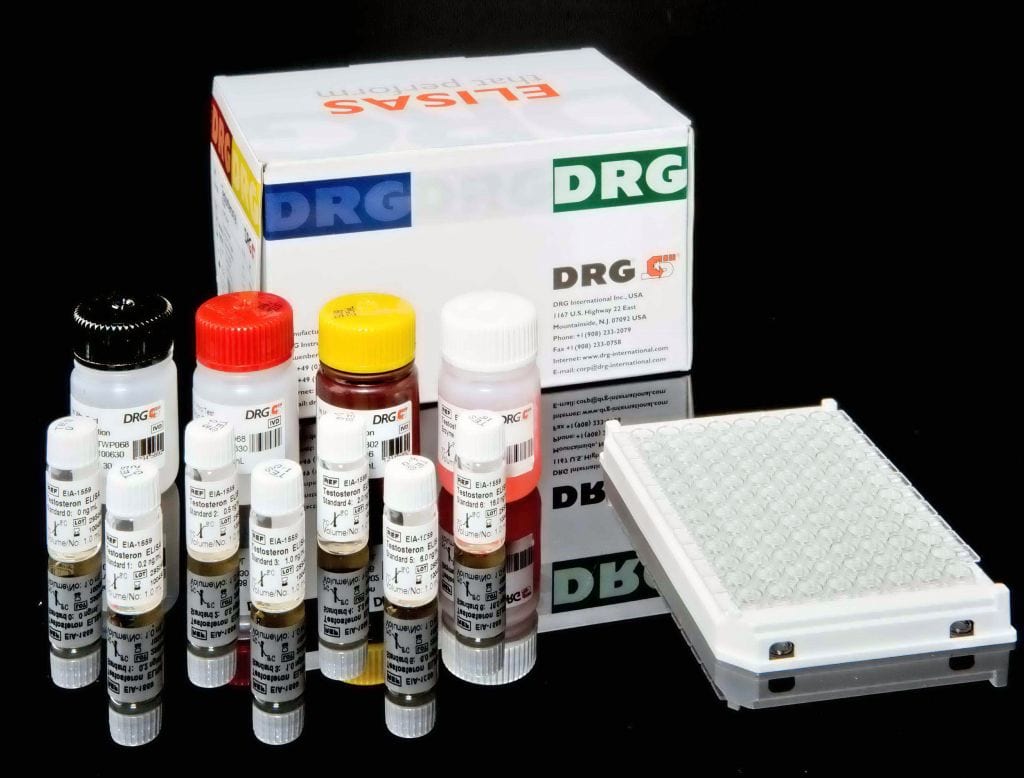
An enzyme immunoassay for the quantitative determination of caridac specific troponin I in serum.
Troponin is the inhibitory or contractile regulating protein complex of striated muscle. It is located periodically along the thin filament of the muscle and consists of three distinct proteins: troponin I, troponin C and troponin T.1-5 Likewise, the troponin I subunit exists in three separate isoforms; two in fast-twitch and slow-twitch skeletal muscle fibers, and one in cardiac muscle.6-8 The cardiac isoform (cTnI) is about 40% dissimilar, has a molecular weight of 22,500 daltons, and has 31 additional amino acid residues that are not present on the skeletal isoforms. 3-4,8-12 Antibodies made against this cardiac isoform are immunologically different from antibodies made against the other two skeletal isoforms,10,13 and the unique isoform and tissue specificity of cardiac troponin I is the basis for its use as an aid in the diagnosis of acute myocardial infarction (AMI).2-4,8-9,13-17 Cardiac troponin I
(cTnI) has been useful in the differential diagnosis of patients presenting to Emergency Departments (ED) with chest pain. 18-20Myocardial infarction is diagnosed when blood levels of sensitive and specific biomarkers, such as cardiac troponin, the MB isoenzyme of creatine kinase (CK-MB), and myoglobin, are increased in a clinical setting of acute ischemia.21-22,23 The most recently described and preferred biomarker for myocardial damage is cardiac troponin (I or T)23. The cardiac troponins exhibit myocardial tissue specificity and high sensitivity. Likewise, cardiac TnI and CK-MB have similar release patterns (4-6 hours after the onset of pain), but the level of cTnI remains elevated for a much longer period of time (6-10 days), thus providing for a longer window of detection of cardiac injury.23-24 Normal levels of cTn I in the blood are very low. After the onset of an AMI, cTnI levels increase substantially and are measurable in serum within 4 to 6 hours, with peak concentrations reached in approximately 12 to 24 hours after infarction.24-28The fact that cTnI remains elevated in serum for a much longer period of time, added to its enhanced diagnostic sensitivity and cardiac specificity, allows for the detection of AMI much earlier after the onset of ischemia (4 hours),1,25 as well as the diagnosis of peri-operative infarction in situations where a high serum level of skeletal muscle proteins are expected.17Additionally,
recent data have identified a measurable relationship between cardiac troponin levels and long-term outcome after an episode of chest discomfort.24,29 The studies suggest that the use of the cTnI demonstrates high predictive value in delineating the high risk group of unstable angina patients,30 and that these tests may be particularly useful in evaluating patient condition prior to discharge from the ED.25,29,31The cTnI Enzyme Imunoassay provides a rapid, sensitive, and reliable assay for the quantitative measurement of cardiac-specific troponin I. The antibodies developed for the test will determine a minimal concentration of 1.0 ng/mL, and there is no ccoss-reactivity with human cardiac troponin T or skeletal troponin T or I.
The cTnI ELISA test is based on the principle of a solid phase enzyme-linked immunosorbent assay. The assay system utilizes four unique monoclonal antibodies directed against distinct antigenic determinants on the molecule. Three mouse monoclonal anti-troponin I antibodies are used for solid phase immobilization (on the microtiter wells). The fourth antibody is in the antibody-enzyme (horseradish peroxidase) conjugate solution. The test sample is allowed to react simultaneously with the four antibodies, resulting in the troponin I molecules being sandwiched between the solid phase and enzyme-linked antibodies. After a 90-minute incubation at room temperature, the wells are washed with water to remove unbound-labeled antibodies. A solution of tetramethylbenzidine (TMB) Reagent is added and incubated for 20 minutes, resulting in the development of a blue color. The color development is stopped with the addition of 1N hydrochloric acid (HCl) changing the color to yellow. The concentration of troponin I is directly proportional to the color intensity of the test sample. Absorbance is measured spectrophotometrically at 450 nm.


