Description
Retinol binding protein (RBP) is from a family of structurally related proteins that bind small hy_drophobic molecules such as bile pigments, steroids, odorants, etc1. RBP is a 21 kDa highly con_served, single-chain glycoprotein, consisting of 182 amino acids with 3 disulfide bonds, that has a hydrophobic pocket which binds retinol (vitamin A). RBP binds retinol in a 1:1 stoichiometry, which serves to not only solubilize retinol but also protect it from oxidation. When in serum, the majority of RBP bound with retinol is reversibly complexed
with transthyretin (prealbumin)2,3. This complex then transports retinol to specific receptors of various tissues in the body. Vitamin A status is reflected by serum concentration as it is hemostatically controlled and does not fall until stores are dramatically reduced4,5. RBP has also been shown to be a useful marker for renal function6 as it is totally filtered by the
glomeruli and reabsorbed by proximal tubules7. This has made urinary RBP (uRBP) a tool to study renal function in heart8 or kidney9 transplant recipients, type 1 and 2 diabetics10, and in people exposed to uranium from mining operations11. Measurement of uRBP levels has also been useful in detection and characterization of diseases including hypertension12 and certain cancers13,14.
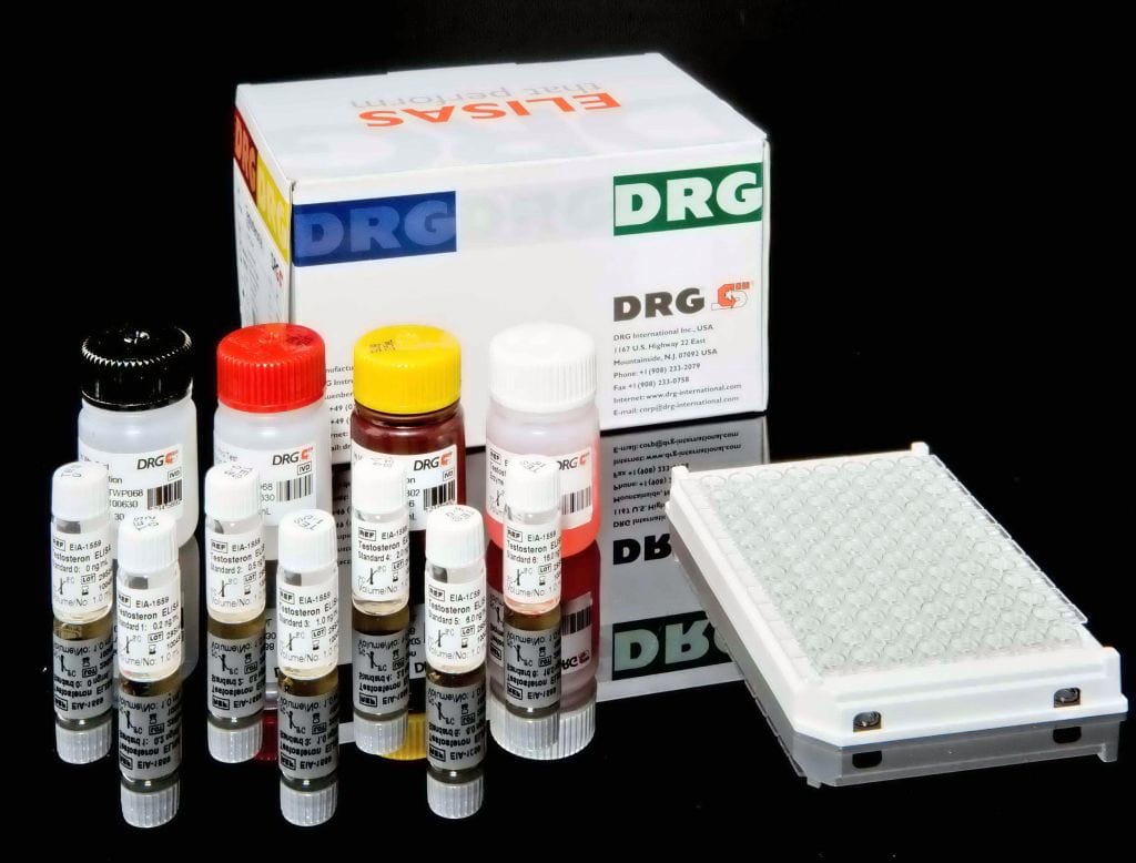
The Urinary Retinol Binding Protein (RBP) kit is designed to quantitatively measure RBP present in urine samples. Please read the complete kit insert before performing this assay. A RBP standard is provided to generate a standard curve for the assay and all samples should be read off the standard curve. Standards or diluted samples are pipetted into a clear microtiter plate coat_ed with an antibody to capture rabbit antibodies. A RBP-peroxidase conjugate is added to the standards and samples in the wells. The binding reaction is initiated by the addition of the RBP polyclonal antibody to each well. After an hour incubation the plate is washed and substrate is added. The substrate reacts with the bound RBP-peroxidase conjugate. After a short incubation, the reaction is stopped and the intensity of the generated color is detected in a microtiter plate reader capable of measuring 450 nm wavelength. The concentration of the RBP in the sample is calculated, after making a suitable correction for the dilution of the sample, using software avail_able with most plate readers.


